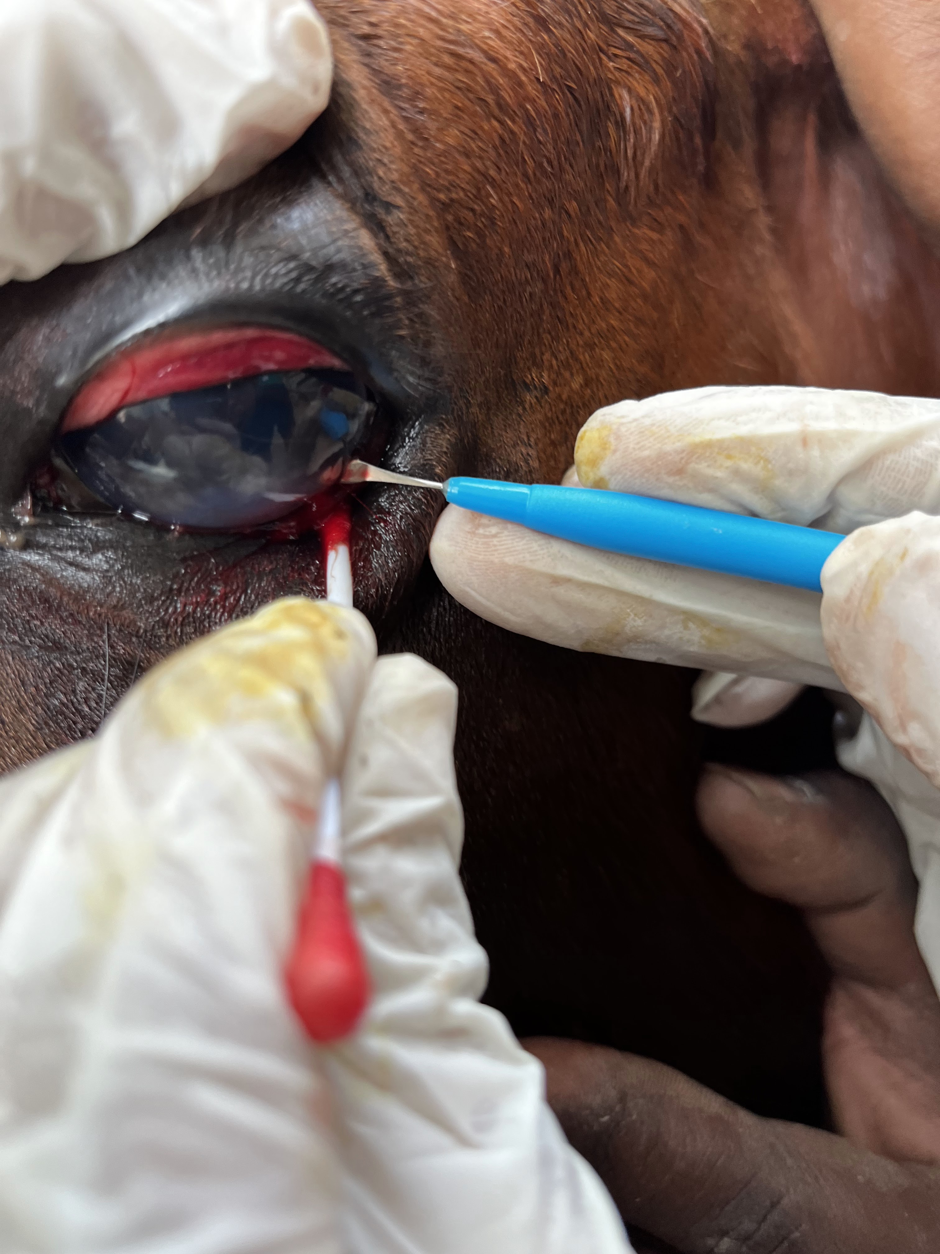
Gunda was 3-year-old stallion that presented with a complaint of corneal opacity in the left eye. Initial symptomatic treatments by the primary attending veterinarian outside of Mysore proved unsuccessful. A detailed ocular examination by a close friend of mine revealed presence of an eye worm inside the anterior chamber. We planned to remove the worm surgically. the horse was sedated using Xylazine and Butorphanol given IV. It was dosed at 0.5mg/kg and 0.02 mg/kg respectively.
Further, the supraortibal nerve and auriculopalpebral nerve were blocked using 2% Lignocaine. The supraorbital nerve was blocked at the level of the foramen to desensitize the supraorbital nerve. the auriculopalpebral nerve was blocked at all the 3 possible sites although just blocking it at one location would have been sufficient.
The limbus was penetrated using a 2.8 mm keratome. About 1 ml of HPMC Viscoelastic material was injected into the anterior chamber to immobilize the microfilaria. A capsulorhexis forceps was then used to extrude the worm outside. the limbal incision was closed using two interrupted sutures of Vicryl 8-0.
The pony was kept on Topical Moxifloxacin which was administered every 3 hours for 10 days. No complications were reported during the recovery. The worm was sent to the Department of Parasitology, Veterinary College, Hebbal for identification. It was identified as Setaria digitata, vector-borne nematode whose prediliction site is the peritoneum. The worm ended up in the eye due to an aberrant migration pattern.
There is a report of medical management of this condition in Sri Lanka. 2 ponies were given a single dose of ivermectin which resulted in the death of these worms but that led to an inflammatory reaction and subsequent uveitis which had to be managed symptomatically. The condition did resolve in the end but took a prolonged course. Majority of articles warrant surgical management for a better outcome. The keratotomy wound should be preferentially closed with 8-0/9-0 Vicryl but some surgeons leave it to heal by secondary intention.


2. Lumb and Jones Veterinary Anesthesia and Analgesia.


Comments
Post a Comment