Animal bites in dogs, cats and in
large animals are one of the most common presentations a veterinarian receives
in his practice. Here's a case of a three-year old Indie, mother of two
puppies, surviving a leopard attack and finding a safer home and a loving
caretaker two weeks later.
'Jenny' was deployed as a ‘guard dog’
at a farm about 70km away from Bangalore. The onlooker who initially reported
the incident to the farm owner witnessed the leopard going for the kill late
one evening. The spikes in Jenny's collar belt and stones which were pelted were
most likely what gave her a second chance to live.
She was prescribed a topical spray, given first aid and prescribed oral antibiotics by a local veterinarian. The owners noticed 'blood' and discharge from the wounds despite the treatment and presented it to us at Prakruthi Vet Hospital for a detailed examination. Jenny was understandingly slightly anemic due to the possible blood loss. Cellulitis, sero-purulent discharge with a foul odour was observed from the wound with fresh maggots. The wound was clearly infected and infested with maggots. Routine treatment was carried out which included thoroughly cleaning, debriding, and flushing the wound and surrounding areas, maggoticidal spray and injectable antibiotics were prescribed.


Jenny got better the very next day and started feeding orally but the owner noticed food and water ingested seeping out of the bite wound and presented her two days later . Suspecting an oesophageal tear, which obviously is the first thing that comes to mind, a contrast esophagogram was performed after feeding barium slurry to detect an oesophageal wall defect if at all present. Radiography of the lateral esophagus did reveal traces of barium outside the usual course of esophagus hinting towards a possible perforation. Assuming the tear was a minor one and hoping it would heal naturally because she was otherwise clinically normal, having food orally and ironically even lunging at cats at the hospital! I deferred surgical exploration/ correction for a couple of days to spare her from another stressful event hoping she would recover. Food and water continued to trickle down the side of the neck whenever fed, which pushed us to take Jenny into surgery.

Operating on the neck isn’t something we do routinely as vets, but is one of the many ‘peaks’ you’ve got to conquer as a ‘Veterinary Surgeon.’ It’s a test of confidence, technical and surgical skills, something which gives a naïve vet the heebie-jeebies. Suddenly, you wished you’d paid more attention while dissecting in anatomy class!
Jenny was sedated with Dexmedetomidine and Butorphanol, induced with Propofol and maintained on Isoflurane through endotracheal intubation throughout the surgery. A gastric tube was also passed to empty gastric contents and for easy identification of the esophagus. A ventral cervical midline approach to the esophagus was preferred over the more complex lateral approach. After incision of the skin and subcutaneous fascia, the sternohyoideus muscles of either side were separated bluntly at the level of pharynx. Careful blunt dissection of the slightly laterally placed (in relation to the midline/trachea) sternothyroideus muscle fascia revealed the esophagus confirmed by the visualization of the gastric tube through the tear. Muscular fascia attached to the esophagus was also carefully separated aborally, later retracting the carotid sheath laterally to the right, guarding the transversely running branch of the carotid artery, the cranial thyroid artery coursing through the sternothyroideus muscle, to the thyroid gland and retracting the trachea to the right. On further exploration of the course of the esophagus, one transverse tear and a longitudinal tear on the dorsolateral surface of the esophagus was confirmed. The transverse tear was initially addressed, suturing it in two layers using Polydiaxanone (PDS) 3-0 in interrupted pattern. First, the mucosa and submucosa were included, and the knots were buried and subsequently, the muscularis and tunica adventitia was sutured with knots outside. The more orally located, larger irregular tear was more challenging to visualize and suture for it was on the dorsal side and we had taken a ventral approach. This defect was sutured blindly in a two-layered continuous pattern with PDS 3-0 owing to incomplete visualization of the tear. The wound was thoroughly flushed with saline and the fistulous tract through which the ingesta was leaking, was also flushed and debrided. The muscles on both the lateral sides which were separated during dissection were opposed using PDS 2-0 in two layers followed by closure of subcutis and skin in a routine manner.


Jenny was prescribed
intravenous Ceftriaxone for five days post-surgery and Pantoprazole for the
three days following surgery during which no oral food or liquids were given. A makeshift,
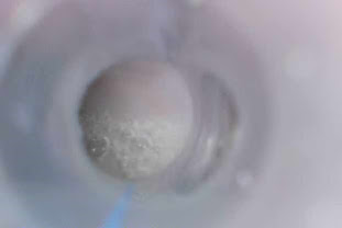
non-surgical endoscope camera which although inferior in quality, having poor
white balance and devoid of an insufflator but which captures clinically significant
and diagnostic images was used for esophagoscopy on the fourth postoperative
day under anesthesia to evaluate mucosal healing. The endoscopic examination
revealed erythema of the esophageal mucosa, suggestive of incomplete / second
intention healing and at another location, a possible perforation of the
esophageal wall although inconclusive owing to the poor quality of the image. The
surgical wound on the other hand was healing well with no discharge from either
the bite wound or the surgical wound which was possible evidence that there was
no esophageal tear. To err on the safer side, the owner was advised to withhold
food for another couple of days followed by which, small quantity of kibbles was
fed at frequent intervals. A second endoscopic examination 3 days after the
first (7th post-op day) revealed complete healing of the esophageal wall.


General Surgical Principles
Esophageal surgery is historically associated with a
higher prevalence of
incisional dehiscence than surgery on other portions of the GIT
because of lack of serosa, segmental
nature of the blood supply, the lack of omental covering, constant motion
caused by swallowing and respiration, and tension at the
surgical site. In the abdomen,
the serosa is
credited with assisting healing of
viscera by the elaboration of a fibrin seal soon after surgery
and by providing a source of pluripotential mesothelial
cells, which is devoid in the cervical esophagus. Monofilament,
minimally reactive, slowly absorbable suture materials,
such as polydioxanone are often preferred for
closure of esophageal incisions considering their healing
potential.
Traditionally,
a two-layer closure technique is used for
the suturing of the esophagus, consisting of a first layer incorporating the
mucosa or submucosa
with placement of the knots in the esophageal lumen and a
second layer consisting of an inverting pattern in the muscularis.
The submucosa is the functional
suture-holding layer of the esophagus and therefore
should be incorporated in at least one layer of sutures.
An interrupted pattern is generally preferred for esophageal anastomoses
and closures to permit esophageal dilatation and to avoid potential interference with the intramural blood supply.
Recommendations on
the time period for withholding per os food and water vary
between 24 hours and 7 days, depending on the type of
esophageal surgery.
Conclusion
1. Always
consider exploring a penetrating wound, no matter where they are located on the
body.
2. Contrast
esophagogram – a valuable diagnostic tool in esophageal disorders.
3. Tears
cranial to or at the level of pharynx are challenging to approach and repair.
4. The importance of Surgical anatomy cannot be over-emphasized, you’ve just got to know it before you cut it!! While operating on the esophagus, take special care to isolate the carotid sheath, cranial and caudal thyroid arteries, and the recurrent laryngeal nerve.
Endoscopy can be a valuable diagnostic to evaluate esophageal healing which in this case could have been done prior to the surgery too. The downside being it requires sedation or anesthesia.
C Finally, credit to the team of PVH and the caretaker for taking excellent care of Jenny post surgery and for providing it a safe home.
jn
en


Comments
Post a Comment