Dermoid
cysts are complex congenital cysts that form long before
birth. Dermoid cysts occur when ectodermal tissue i.e., skin, and
adnexal skin structures collect under the skin during fetal development. These
cysts may contain hair, teeth, or even nerves. They usually appear at birth and
grow very slowly or usually remain the same size.
Branchial cleft cysts
are similar pathologies when the tissues on either one or both sides of the
neck develop abnormally. These can sometimes, but need not necessarily communicate
with the skin through an opening called branchial cleft sinus tract.
Dermoid cysts are commonly
seen in the eyes of dogs, but this was the first case where I found a similar
pathology located internally. A 3-year-old Labrador had a soft fluctuating lemon-sized
swelling ventral to the right submandibular lymph node. It was clinically insignificant
during previous visits and an attempt to aspirate the contents turned out to be
futile. The content was a brownish liquid with visible fat globules. 5 months
later, the dog was presented with a complaint of scratching at the site.


Surgical exploration and
excision was planned. Although the fluctuating swelling was palpable and visible
as a bulging eminence over the skin, it was deeply situated and required
careful dissection around vital neurovascular structures of the neck to fully
isolate the cyst and excise it.

.jpeg)
Dissection of the cyst
after the surgery revealed similar fluid to that obtained through aspiration. A
layer of skin with hair follicles was found loosely attached to the inner layer
of the cyst which could easily be separated. A section of the same was sent for
histopathology.


The histopathology report
stated “Section studied shows tissue fragments lined by stratified squamous
epithelium with separately lying fragments of keratin debris. Adnexal structures
are noted in the wall. The section is negative for granuloma or malignancy.
Features are suggestive of dermoid cyst.”
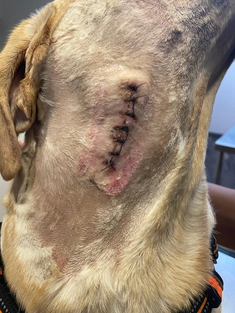

Images showing the healing of surgical wound 10 days post-surgery and after suture removal
I couldn’t find many clearly defined and published reports of either condition in dogs and hence is difficult to differentiate between the two cysts. However, considering the location of the cyst and the histopathology report, I believe what I excised was a branchial cyst. Complete surgical excision is the only foolproof modality advised for the treatment of branchial cysts.


Comments
Post a Comment