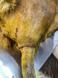
A seven-year-old male Siberian Husky was presented with a complaint of an ulcerating mass on the dorsolateral region of the base of the tail. The mass persisted for more than six months but started to ulcerate a week before.
A tentative diagnosis of a supra caudal gland tumor/ tail gland tumor was made taking into account the tumor's location and the case's signalment.
The large size of the tumor and the 2cm margins that had to be attained during surgical removal meant that a large area had to be prepped and required either a tension-relieving skin closure technique or a subdermal plexus flap. In order to avoid these relatively extensive and invasive procedures, a castration was planned and performed hoping the size of the mass would decrease once there was no influence of Testosterone. The castration did not help and a decision was made to excise the mass.
Primary skin closure was impossible due to the large skin defect that was created. A subdermal plexus advancement flap was created over the rump, advanced caudally, and apposed using Polyglecaperone-25 in a simple interrupted pattern.
An active suction drain would have helped counter the seroma owing to a lot of dead space but wasn't placed due to a lack of availability during the surgery. Nonetheless, despite considerable serous discharge after the surgery, the wound healed without complications, and the sutures were removed 21 days after the surgery.
Preoperative images of the tumor at the base of the tail
Intraoperative image showing the large skin defect after excision of the tumor
Postoperative images showing the closure of the skin defect after caudal advancement of a subdermal plexus flap
7th Day Post-operative day images of the surgical wound showing the intact sutures and desiccation of the serous discharge leading to the scabs on the wound
21st Day Post-operative day images of the surgical wound after suture removal


Comments
Post a Comment